
Researchers explore link between medical imaging and cancer risk in children

Study of nearly 4 million children finds that 10% of pediatric blood and bone marrow cancers may have stemmed from radiation exposure
A groundbreaking study has concluded that radiation from medical imaging, even at relatively low doses, is associated with a higher risk of blood cancers in children.
The researchers examined data from nearly 4 million children and adolescents and estimated that 1 in 10 of the children’s blood cancers — some 3,000 cancers in all — may be attributable to radiation exposure from medical imaging. The risk increased proportionally to the cumulative amount of radiation the children received.
“This study provides robust, directly observed evidence of a clear dose-response relationship between radiation from medical imaging and hematologic malignancy risk in children and adolescents,” said co-senior author Diana Miglioretti, PhD, an affiliate investigator with Kaiser Permanente Washington Health Research Institute (KPWHRI) and professor and chief of the Division of Biostatistics at University of California Davis Health.
The study, which was funded by the National Institutes of Health, analyzed data from 6 health systems in the United States and Ontario, Canada, including Kaiser Permanente Washington. It was published in The New England Journal of Medicine.
The investigation is the first comprehensive assessment using data from children in North America that quantifies the association between radiation exposure from medical imaging and blood and bone marrow cancers, such as leukemia and lymphoma, which are the most common forms of cancer in children and adolescents.
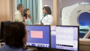
Medical imaging saves lives by enabling timely diagnosis and effective treatment, but it also exposes patients to ionizing radiation, a known carcinogen, particularly through computed tomography (CT). The authors caution that doctors and parents should avoid excessive radiation doses and minimize exposure when clinically feasible.
“Children are particularly vulnerable to radiation-induced cancer due to their heightened radiosensitivity and longer life expectancy,” said Rebecca Smith-Bindman, MD, a radiologist and professor of epidemiology and biostatistics, as well as obstetrics, gynecology, and reproductive sciences at University of California San Francisco, and the first author of the paper.
“While medical imaging can be lifesaving, our findings underscore the critical need to carefully evaluate and minimize radiation exposure during pediatric imaging to safeguard children’s long-term health,” said Smith-Bindman, who is also a member of the Philip R. Lee Institute for Health Policy Studies. “This involves ensuring that imaging is performed only when it provides essential information for the child’s care and, in cases such as CT scans, using the lowest possible radiation doses.”
Documenting risks in children
The study used a retrospective cohort design, looking back at the complete imaging histories of 3.7 million children who were born between 1996 and 2016. It found that the most frequently performed CT scan was of the head and brain, while chest radiography was the most common imaging exam overall.
Investigators found a significant relationship between cumulative radiation dose and the risk of a hematologic malignancy, which includes tumors affecting the blood, bone marrow, lymph, and lymphatic system.
The risk of developing cancer varied significantly by imaging modality. CT — which is used to detect many abnormalities such as tumors, heart disease, and injuries of the spinal cord and brain — entails significant radiation exposure. But radiographs — which are used to diagnose both broken bones and pneumonia — expose children to much lower doses.
For children who underwent a head CT, the researchers attributed about a quarter of the children’s subsequent hematologic malignancies to radiation exposure. For those who had radiographs, by contrast, they estimated that only a small fraction of the children’s subsequent cancers were associated with radiation exposure.
Getting one or two head CTs was associated with a 1.8-fold increased risk of a cancer diagnosis, and this rose to 3.5 times for children who received more scans and were therefore exposed to more radiation.
Altogether, 2,961 hematologic malignancies were diagnosed during the study period. Lymphoid malignancies accounted for 79.3% while myeloid malignancies and acute leukemia together accounted for 15.5%. About 58% of cancers occurred in males, and about half were diagnosed in children under age 5.
The authors said that up to 10% of hematologic malignancies in children could be prevented by reducing unnecessary imaging and optimizing radiation doses. In many cases, the authors said, substituting non-ionizing imaging modalities like ultrasound or MRI may be feasible without compromising diagnostic accuracy.
Weighing benefits and risks
The authors emphasized that while medical imaging remains an invaluable tool in pediatric care, their findings highlight the need to carefully balance its diagnostic benefits with potential long-term risks.
“Our findings align with international research highlighting that children are especially radiosensitive,” Miglioretti said. “It’s crucial for clinicians to weigh the immediate benefits of imaging against potential long-term health risks and to optimize imaging protocols to minimize radiation exposure.”
The research was supported by the National Cancer Institute of the National Institutes of Health. The Ontario, Canada, portion of the study was supported by ICES, which is funded by an annual grant from the Ontario Ministry of Health and Long-Term Care.
Erin Bowles, MPH, KPWHRI’s director of Collaborative Science, was a coauthor on this paper.
This story was adapted from a news release from University of California San Francisco.

Featured
Researcher
Erin J Bowles, MPH
Director, Collaborative Science
Access researcher directory
Research
Medical imaging rates keep rising despite push to reduce them
Potential harms should be considered before scanning, conclude UC and Kaiser Permanente researchers.
Research

Supporting young adults through cancer and beyond
A potential new care model for young cancer survivors centers patient needs, support networks.
VOICE STUDY

Understanding young adults’ experiences with cancer
The VOICE study aims to improve the health and health care of people who had cancer as adolescents and young adults.